
Our Approach · The Legends · Era III
Era III · b. 1922 · Lubbock, Texas
William G. Blair
The Cartographer
William G. Blair (born 1922) built upper cervical chiropractic around each patient's own bone. He opened his Lubbock practice on December 24, 1949. In 1964 Blair reported that the joints between skull, atlas, and axis are asymmetric in most people, and he designed radiographs that image those joints as they are formed in that person. The Blair Upper Cervical Technique computes the adjusting angle and the torque from that anatomy. The Unified Model of Tone holds that specificity is correspondence to the individual, which is what Blair measured.
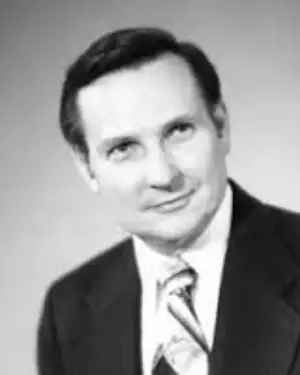
Born
July 30, 1922 · Pemeta, Oklahoma
Practice
Lubbock, Texas · opened December 24, 1949
Published
Blair Upper Cervical Spinographic Research · November 1964
Taught
Primary and Advanced seminars · about 1961 to 1980
Founded
The Blair Upper Cervical Technique
Pemeta to Lubbock
Blair came to chiropractic as a patient whose asthma nothing had touched
William George Blair was born on July 30, 1922 in Pemeta, Oklahoma, and was diagnosed with bronchial asthma at nine months old. His parents tried every treatment available to them, chiropractic among them, and none of it held. Blair still finished high school in 1940 in the top ten percent of his class, having missed a great deal of it to illness. He spent two years at Oklahoma A and M College, then moved to Lubbock, Texas, where the dry climate suited his lungs and family was close.
The referral that ended a sales career
Blair was working as a salesman when his wife introduced him to upper cervical chiropractic. The two had married on July 20, 1946. A family friend referred him to Dr. Leon Halstead in Houston. Blair was examined, adjusted, and sent home with an account of what to expect. His breathing eased almost at once. Blair decided to become a chiropractor, packed up his family, and moved to Davenport, Iowa.
Davenport gave Blair the method he would later correct
Blair trained at the Palmer School of Chiropractic in the Upper Cervical Specific tradition, the X-ray guided Hole In One work of B.J. Palmer. He returned to Lubbock, rented a small building with an office in front and an apartment behind it, and opened on December 24, 1949. That clinic is still seeing patients past its seventy fifth year. Everything Blair later built began as a correction to the analysis he was taught in Davenport.
The uneven result
Blair went looking in the bone for the reason his results varied
Blair's Upper Cervical Specific results varied more than the method predicted. Patients adjusted by the same hands from the same films improved by very different amounts. Blair traced the variation to the analysis rather than to the delivery. He suspected that osseous asymmetry, which he called malformation, was corrupting the listing the film produced. A listing computed from a distorted picture sends an accurate thrust to the wrong place.
Asymmetry enters the analysis as error
Conventional upper cervical views read misalignment against an assumed mirror image neck. Blair held that the assumption fails in most people. Where a patient's two sides differ, part of what the film shows as displacement is that patient's own architecture. E.A. Addington wrote the technique description for the Council on Chiropractic Practice in October 1995. He records the concern in Blair's own terms: the effect of asymmetry on the accuracy of the spinographic analysis in producing a valid adjustive listing. Blair concluded that atlases were being adjusted that did not need it.
Skeletal specimens told Blair the atlas cannot slide sideways
Blair studied skeletal specimens, one of them from B.J. Palmer's osteological collection, and drew two conclusions about how the atlas can move. The slope of the occipital condyles and of the atlas lateral masses forms an osseous lock, so the atlas cannot travel in a truly lateral direction. The condyles and the lateral masses are also complementary in shape, so the atlas cannot rotate against the occiput in the coronal plane without gapping the joint. Misalignment therefore has to run obliquely, along the joint's own planes.
The 1964 count
Blair counted asymmetry in hundreds of skulls and published the frequencies in 1964
Blair published A Synopsis of the Blair Upper Cervical Spinographic Research in November 1964, in the first issue of Science Review of Chiropractic, pages 1 to 19. The work rested on hundreds of cranio-atlanto-axial spinographs. Blair reported how often the structures used in standard cervical analysis are built asymmetrically, and the frequencies were not marginal.
The frequencies Blair reported
An anterior occipital condyle appeared in 79 percent of cases. A foramen magnum whose anterior to posterior plane failed to parallel the sagittal plane of the skull appeared in 77 percent. A short occipital condyle appeared in 77 percent of cases measured against the orbital floor, 64 percent against the base line of the skull, and 66 percent against the vertical line. The right condyle measured narrower than the left in 83 percent. Blair called such malformations the rule rather than the exception.
Convergence angle measurements put numbers on the individual case
Addington and colleagues plotted the atlanto-occipital convergence angle in 45 cases and reported the work at Life Chiropractic College in December 1986. Pooled across both sides the mean angle came to 24.3 degrees, with sample values running from 6 to 58 degrees. Inside a single person the left and right convergence angles differed by an average of 7.25 degrees, and in one case by 26.5 degrees. The two angles correlated weakly within a subject, at r equal to .40. A standard reference describes the average of a population whose halves do not match each other.
Anatomists reached Blair's conclusion from their own specimens
Gawlikowska-Sroka and colleagues measured the occipital condyles of 112 dry human crania from the ROMA skeletal collection in Rome and published in Folia Morphologica in 2026 (PMID 42052736). Sexual dimorphism came out below 5.5 percent on every parameter, and age explained almost nothing. What the series showed was pronounced individual variability, with bilateral asymmetry expressed mainly as fluctuating asymmetry. The authors closed by urging individualized anatomical assessment of the craniovertebral junction. Blair had built a clinical system on that conclusion sixty two years earlier. The atlas carries its own variants: a 2026 systematic review put the arcuate foramen, a bony bridge over the vertebral artery groove, at roughly 3 to 15 percent of the population (PMID 41297626).
In reality, one of our greatest constants is the presence of non-symmetry in the bilateral formation of these structures.
William G. Blair, A Synopsis of the Blair Upper Cervical Spinographic Research, Science Review of Chiropractic, November 1964The Blair Principle
The Blair Principle states that the atlas misaligns obliquely, along its own joint planes
Blair set the principle out in his 1968 Ph.C. thesis at Palmer College of Chiropractic. In an atlas that is neither fractured nor dislocated, misalignment against the occipital condyles runs obliquely anterior or obliquely posterior, parallel to one or both of the atlanto-occipital convergence angles. The consequence for treatment is immediate. If a misalignment travels along the joint's own long axis, the correction has to travel there too, and that axis is a measurement in each patient.
The surfaces of one joint match each other even when the two sides do not
One simplifying assumption carries the whole Blair analysis. Two bones that form an articulation develop so that their margins are identical in conformation, whatever the asymmetry elsewhere in the skull. A properly imaged, properly aligned articulation therefore shows a recognizable relationship at the osseous margins. Blair drew a further conclusion from it. A vertebral misalignment, absent fracture, has to occur at an articulation, which makes the articulation the most anatomically accurate place to assess where a vertebra sits.
The two joints Blair works move unlike anything below them
Panjabi, Dvorak, and colleagues measured three dimensional movement in the upper cervical spine and published in Spine in 1988 (PMID 3194778). At the occiput to atlas joint they recorded 3.5 degrees of flexion, 21.0 degrees of extension, 5.5 degrees of lateral bending, and 7.2 degrees of axial rotation to a side. The atlas to axis joint gave 38.9 degrees of axial rotation to a side, the largest single motion anywhere in the spine. Neither joint carries a disc, so bone shape governs the movement more completely here than anywhere else in the column.
The imaging series
Blair built an imaging series that photographs the patient's own joint planes
The Blair protocol runs in two stages, and the first exists to set up the second. A scout series of base posterior, A-P open mouth, and five degree rotated lateral cervical views establishes the angles from which this patient's atlanto-occipital and apophyseal joints can be seen edge on. The base posterior view carries the most weight, since the convergence angle of each atlanto-occipital joint is plotted there. It also shows the asymmetry of the foramen magnum, condyles, lateral masses, and spinal canal.
What a protractoview shows that a standard film cannot
The articular series makes the pictures the analysis is read from. The patient is turned until the angle of the chair equals the convergence angle of the joint under study, so the central ray runs along that joint's own long axis. On a conventional open mouth or lateral film the two sides stack on each other and the joint margins hide behind overlying bone. A protractoview separates them and images the anterolateral margins cleanly. The Blair Protractoclamp, a patented positioning device, holds the patient at the exact degree of rotation the view requires.
Three readings at the joint margin, and one adjusting formula out of them
Each margin receives one of three readings. Juxtaposed means the two margins line up. Overlapped means the atlas lateral mass margin sits further anterolateral than the condyle margin, and underlapped means it sits less far. Readings from both sides combine into the misalignment pathway, the listing, and the adjusting formula. Addington and Muncy estimated in 1989 that at least 578,782 distinct upper cervical adjustments exist inside the Blair system, counting only C1 through C4.
Cone beam CT now returns the same geometry in about thirty seconds
Most Blair Upper Cervical offices image with cone beam computed tomography and have for more than a decade. One rotation around the head returns a volume the doctor re-slices along any plane afterward, which dissolves the positioning problem the Protractoclamp was built to solve. The analytic question is unchanged. Where does this joint's margin sit, on this side, in this person. Blair's requirement was that no vector be derived from anything except the patient's own joint.
The torque Blair kept
Blair kept B.J. Palmer's torque and made its direction an output of the analysis
The Blair Toggle-Torque was developed from B.J. Palmer's own adjusting. Blair learned the thrust in Davenport from watching Palmer work, and the technique's own account is explicit that the move came out of Palmer's hands. What Blair carried forward from it was the torque. Addington's 1995 description states the rule plainly. Superior or inferior torque is used, depending on the misalignment and on the contact the doctor chooses. Torque direction in Blair Upper Cervical is a computed output of the film.
Palmer described torque as a corkscrew moving in three directions
Palmer's 1934 text describes a torque adjustment as a corkscrew motion combining three directions at once: superior or inferior, circular, and obliquely upward or downward. His reasoning was symmetrical. A subluxation that arrived through a three direction twist has to be corrected through one. H.I.O. Knee Chest carries that thrust with recoil, delivered with the patient kneeling and the chest and face down.
The successors dropped the torque or standardized it away
The precision upper cervical systems that grew out of John F. Grostic's work read a lateral, a nasium, and a vertex film, and measure how far the atlas departs from a right angle reference. NUCCA replaced Palmer's toggle with a measured low force contact taken in side posture. Atlas Orthogonal replaced the hand with a percussion instrument. Neither delivers a hand torque whose direction is derived for the patient in front of the doctor. Blair systematized the torque back in and tied its direction to the listing.
The pisiform lead is what makes the torque repeatable
The patient lies in side posture with the head on a drop headpiece. The thrust is a toggle without recoil carrying 180 degrees of torque. Its defining mechanic is the pisiform lead, in which the contacting surface of the doctor's pisiform stays in firm contact with the segmental contact point throughout the thrust rather than describing a helical pathway. One listing breaks the rule. A posterior-inferior atlas takes no torque, and there the doctor does recoil the hands from the thrust.
The working force is about five pounds
Thomas Forest, an author of the 2010 reliability study, puts the working force of a Blair adjustment at roughly five pounds, about what it takes to crush a cherry tomato. Nothing is rotated or stretched, and no cavitation is sought. The drop headpiece falls about a quarter of an inch, and that startle is usually the loudest part of the visit.
Two findings before any adjustment
Blair required a heat pattern and a leg check before he would adjust
Films describe the misalignment, and Blair did not treat them as permission to adjust. He established nerve interference from two findings taken together: a persistent differential paraspinal dermothermographic pattern in the cervical region, and a functional leg length deficiency. Both had to be present on two occasions no more than seven days apart before Blair would adjust again.
The heat pattern is defined by what repeats
A dual probe instrument glides from about the T1 spinous process up to the superior nuchal line, recording the left to right heat difference continuously. Two readings, preferably three, taken on separate days, establish the patient's characteristic pattern. That pattern is defined by the unilateral heat breaks present on every pre-adjustment reading. A single trace cannot separate a pattern from a moment.
Holding time is the outcome Blair tracked
Blair Upper Cervical counts a correction as held for as long as the characteristic heat pattern stays away, and past the first several weeks the benchmark is measured in weeks rather than days. Testing runs twice weekly through the corrective phase for at least six to eight weeks. A failure to hold is read as a reason to re-examine the case rather than to adjust harder. Holding time reports on how well the analysis matched the patient.
The teaching
Blair taught the method from about 1961 and kept refining it after he stopped
Blair showed the work privately for years before he taught it. Addington records that Blair opened it to the field in about 1961, on the unremitting insistence of a trusted colleague. The curriculum ran as Primary and Advanced courses of three days each, the Primary covering spinographic imaging and misalignment concepts and the Advanced covering adjustive methods. For a time the two ran back to back as a five day course, and Blair dropped that arrangement as too demanding of the students. He taught actively until 1980.
More than three thousand cases went into the refinement
Blair's research covered more than 3,000 cases across his career. The views, the listings, and the thrust were all revised against them. He retired from practice in 1982 and went on developing adjustments for multiple misalignments of the atlas afterward, field tested by R. Weldon Muncy of Lancaster, California. Teaching and research passed to the William G. Blair Chiropractic Society after his death. The Society is a Texas nonprofit that credentials instructors, sponsors research, and holds an annual conference.
The Society made reliability its research program
Hubbard, Vowles, and Forest put 25 examiners in front of 100 protractoviews, 7 chiropractic students and 18 doctors of chiropractic among them. Their study appeared in the Journal of Chiropractic Medicine in 2010 (PMID 21629551). Overall interexaminer agreement reached a kappa of 0.62 and intraexaminer agreement 0.81. Among advanced certified practitioners the values were 0.74 and 0.92. Every value carried P less than .001. A method whose whole claim rests on a reading has to show that the reading transfers between hands.
Subluxated articular misalignments are analyzed as they displace the neural rings, thereby occluding the neural canal and compromising cord space.
E.A. Addington, Overview of the Blair Cervical Technique, Council on Chiropractic Practice, October 1995The dated record
Ten dated facts about William G. Blair, with the source for each
1922. Blair was born on July 30 in Pemeta, Oklahoma, and was diagnosed with bronchial asthma at nine months old.
1940. Blair graduated high school in the top ten percent of his class, then spent two years at Oklahoma A and M College.
1946. Blair married on July 20, and his wife introduced him to upper cervical chiropractic.
1949. Blair opened his Lubbock practice on December 24, after graduating from the Palmer School of Chiropractic and moving his family back from Davenport.
1964. Blair published A Synopsis of the Blair Upper Cervical Spinographic Research in Science Review of Chiropractic, volume 1, issue 1, pages 1 to 19, in November.
1968. Blair set out the Blair Principle of Atlanto-Occipital Misalignment in his Ph.C. thesis at Palmer College. He also ran a four part series, For Evaluation: For Progress, in the International Review of Chiropractic that year.
1961 to 1980. Blair taught Primary and Advanced seminars at locations across the United States, running three days each.
1982. Blair retired from practice and kept developing adjustments for multiple misalignments of the atlas, which R. Weldon Muncy field tested.
1989. Addington and Muncy presented their estimate of at least 578,782 distinct upper cervical adjustments in the Blair system at the Society's fourth annual convention in Kansas City, October 28 to 29.
1995. Addington prepared the full technique description for the Council on Chiropractic Practice at Chandler, Arizona, on October 2 and 3. It remains the standard account of the method.
Blair and the model
Blair made the person the reference, and the Unified Model of Tone builds on that
Blair's rule is that no correction may be derived from anything except this patient's own joint. The Unified Model of Tone reaches the same rule from the other direction. The model separates specificity from amplitude and defines specificity as correspondence between the structure of an input and the structure of the patient. Correspondence belongs to the reading, because an input can only match a pattern that has already been found. Blair located that principle in bone and turned it into a measurement.
Asymmetry is geometry to work with rather than error to erase
An orthogonal analysis treats departure from a right angle as the thing to close. Blair treated the same departure as the information the correction is computed from. The 2026 Rome series supports the reading. Asymmetry of the occipital condyles is predominantly fluctuating asymmetry, which is a signature of one individual's developmental history rather than a population trait. The model holds that tone carries an accumulated history, and a Blair film reads a piece of it directly.
The upper cervical junction is a leverage point, a place where a small input moves the whole system, and it is one of several
Blair named the cervical spine as his exclusive concern and never widened it. The model holds that a body ordinarily carries several points of critical tension at once, each able to reorganize the whole, with their relative leverage shifting from week to week. That position explains why incompatible upper cervical geometries all report the same class of change. NUCCA and Atlas Orthogonal measure the same three inches of anatomy against a reference Blair rejected, and each has sustained a field of practitioners for more than half a century.
What a contact at the atlas actually reaches
Hack and colleagues described a connective tissue bridge running from the rectus capitis posterior minor muscle to the spinal dura, in Spine in 1995 (PMID 8610241). Kahkeshani and Ward reviewed that myodural bridge in Clinical Anatomy in 2012 (PMID 22488993). The suboccipital region is the only place in the body where contractile muscle attaches to spinal dura. Boyd-Clark, Briggs, and Galea counted spindles in the deep cervical muscles and reported the densities in Spine in 2002 (PMID 11923661). A Blair contact lands in dense sensory tissue that is mechanically continuous with the covering of the cord. What the nervous system does with a signal of that quality sits at input quality.
Holding time is a measurement of time course
The model holds that how a system returns to baseline after a demand is a reading of tone in its own right, taken before any clinical outcome is known. Blair built his outcome measure on that quantity. He recorded how long a patient held a correction before the characteristic heat pattern returned, and he treated a shortening hold as information about the case. The library's account of that quantity sits at time course.
Where the model goes past what Blair held
Blair's stated objective was relief of adverse mechanical influence on the neurological structures of the cervical spine, and maximum patency of the neural canal. He set aside biomechanical and orthopedic standards of alignment as goals in their own right. The model names the variable underneath the geometry. Tone is the organizing state the nervous system holds across mechanical tension, neural excitability, autonomic regulation, and sensory gain, readable at every scale and able to deviate in two directions. Tone inside its healthy range is health, because that range keeps the ability to adapt to load. Tone that drifts or distorts outside it is what shows up as illness and disease.
Where Blair sits in this library
Blair Upper Cervical carries the imaging protocol, the listings, and the adjustment in full. B.J. Palmer supplied the thrust Blair reworked and the osteological collection he studied. H.I.O. Knee Chest is the method Blair trained in and corrected. Grostic founded the orthogonal line Blair broke with, and Roy Sweat carried it into instrument delivery. Toftness reaches a comparable force scale from an instrument reading instead of a film. Tone defines what every technique in this library is working on.
Questions people ask
Common questions about William G. Blair
What did William Blair contribute to the understanding of tone?
Blair made the patient the reference for the correction. He showed in 1964 that the joints between skull, atlas, and axis are asymmetric in most people, then built imaging that derives the adjusting angle and the torque from that person's own joint planes. The Unified Model of Tone holds that specificity is correspondence to the individual and that accuracy is decided in the reading. Blair carried that principle down to the level of bone and made it a measurement.
Who was William G. Blair?
William George Blair was an American chiropractor born on July 30, 1922 in Pemeta, Oklahoma. He practiced in Lubbock, Texas from December 24, 1949. Blair had bronchial asthma from nine months old and entered chiropractic as a patient. He graduated from the Palmer School of Chiropractic and published his spinographic research in 1964. Blair founded the Blair Upper Cervical Technique, taught it from about 1961 to 1980, and retired from practice in 1982.
What is the Blair Principle of Atlanto-Occipital Misalignment?
The principle holds that an atlas which is neither fractured nor dislocated misaligns obliquely anterior or obliquely posterior, parallel to one or both of the atlanto-occipital convergence angles. Blair reached it from skeletal specimens. The slope of the condyles and the atlas lateral masses forms an osseous lock that rules out a truly lateral slide, and their complementary shapes rule out coronal rotation without gapping the joint. He set the principle out in his 1968 Ph.C. thesis.
Why did Blair reject the standard upper cervical X-ray views?
Standard views read misalignment against an assumed symmetric neck. Blair counted the assumption and found it false in most people. An anterior occipital condyle appeared in 79 percent of his cases and a right condyle narrower than the left in 83 percent. Where a patient is asymmetric, part of what the film shows as displacement is that patient's own architecture, so the listing derived from it can send an accurate thrust to the wrong place.
What is the torque in a Blair adjustment?
The Blair Toggle-Torque is a toggle without recoil carrying 180 degrees of torque, taken from B.J. Palmer's own adjusting. Superior or inferior torque is used depending on the misalignment and on whether the doctor takes an ipsilateral or contralateral contact, so its direction is computed from the film for that patient. One listing, a posterior-inferior atlas, takes no torque at all, and there the doctor recoils the hands from the thrust.
How is Blair different from NUCCA and Atlas Orthogonal?
All three work the same two bones and all three trace to B.J. Palmer. NUCCA and Atlas Orthogonal descend from John F. Grostic and measure how far the atlas departs from a right angle reference. Blair holds that joint planes differ between a person's two sides and between people, so no standard reference describes them. Blair also lists and adjusts the segments below the atlas, down to C4.
Sources
Sources for this page
- Addington EA. Overview of the Blair Cervical Technique. Prepared for the Council on Chiropractic Practice, Chandler, Arizona, October 2 to 3, 1995. Full text. The history, the objective, the two stage spinographic protocol, the toggle-torque and its torque rule, and the teaching chronology.
- Blair WG. A synopsis of the Blair upper cervical spinographic research. Science Review of Chiropractic 1964;1(1):1-19. The malformation frequencies and the constancy of non-symmetry, reported from hundreds of cranio-atlanto-axial spinographs.
- Blair WG. Blair Upper Cervical Spinographic Research; Primary and Adaptive Malformations; Procedures for Solving Malformation Problems; Blair Principle of Occipito-Atlanto Misalignment. Ph.C. thesis, Palmer College of Chiropractic, 1968.
- Blair WG. For evaluation: for progress. International Review of Chiropractic 1968: February 22(8):8-11; March 22(9):8-11; June 22(12):8-11; August 23(2):8-11.
- Hubbard TA, Vowles BM, Forest T. Inter- and intraexaminer reliability of the Blair protractoview method. J Chiropr Med. 2010;9(2):60-68. PMID 21629551. Kappa 0.62 interexaminer and 0.81 intraexaminer overall, 0.74 and 0.92 among advanced certified practitioners.
- Addington EA, Howard MG, Pruitt MA, Spears CE. Objectivity of Blair atlanto-occipital articular convergence angle measurements. Third Annual Upper Cervical Symposium, Life Chiropractic College, December 5 to 7, 1986. The 45 case convergence data.
- Addington EA, Muncy RW. Estimated number of distinct upper cervical adjustments in Blair technique. Fourth Annual Convention, William G. Blair Chiropractic Society, Kansas City, October 28 to 29, 1989.
- Panjabi M, Dvorak J, Duranceau J, Yamamoto I, Gerber M, Rauschning W, Bueff HU. Three-dimensional movements of the upper cervical spine. Spine. 1988;13(7):726-730. PMID 3194778.
- Gawlikowska-Sroka AK, Gromnicka D, Szczurowski J, Moryś J, Dzięcioł-Anikiej Z, Dzięciołowska-Baran E. Morphology, morphometry and bilateral asymmetry of the occipital condyles in a historical human skeletal collection. Folia Morphol. 2026;85:e01726070. PMID 42052736. 112 crania, pronounced individual variability, asymmetry predominantly fluctuating.
- Hack GD, Koritzer RT, Robinson WL, Hallgren RC, Greenman PE. Anatomic relation between the rectus capitis posterior minor muscle and the dura mater. Spine. 1995;20(23):2484-2486. PMID 8610241.
- Kahkeshani K, Ward PJ. Connection between the spinal dura mater and suboccipital musculature: evidence for the myodural bridge and a route for its dissection. Clin Anat. 2012;25(4):415-422. PMID 22488993.
- Boyd-Clark LC, Briggs CA, Galea MP. Muscle spindle distribution, morphology, and density in longus colli and multifidus muscles of the cervical spine. Spine. 2002;27(7):694-701. PMID 11923661.
- Valenzuela-Fuenzalida JJ, et al. Arcuate foramen of the atlas vertebra and its correlation with clinical implications in the craniocervical region: a systematic review and meta-analysis. World Neurosurg. 2026;205:124652. PMID 41297626.
- About Dr. William Blair. Blair Chiropractic. Biography. Birth date and place, the asthma diagnosis at nine months, the 1940 graduation, Oklahoma A and M, the July 20, 1946 marriage, Dr. Leon Halstead, and the December 24, 1949 opening.
This page is history and education. It is not medical advice, and nothing here is a promise of outcome. Consult a licensed practitioner about your own condition.
